Originally answered on Quora: Which regions of the brain experience the highest levels of oxidation due to aging?
Oxidative stress in our aging brains is all about finding the balance between the natural production of reactive oxygen species and the forces that keep them in check. As we grow older, our brains struggle to maintain the right balance between these harmful molecules and our antioxidant defenses. This can result in damage to our brain cells, reduced ability to think and remember, and even increase the risk of diseases like Alzheimer’s. One proposal states that brain regions acting as “cortical hubs” would experience the highest level of activity and metabolism, and consequently, would also be the most vulnerable to oxidative stress in the aging brain.
What are cortical hubs?
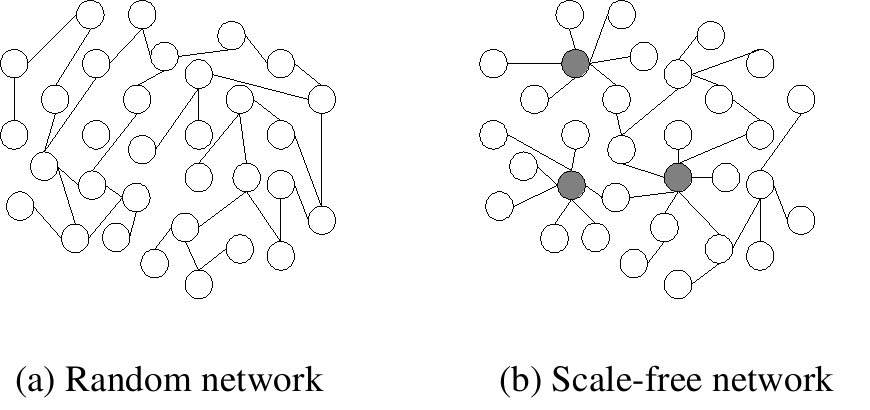
The connectivity architecture of the human brain has been described as a small-world network, following an exponentially truncated power-law distribution (Achard et al., 2005). A key characteristic of this type of network is that while most nodes will have a low degree of connections, a select few will have a dense number of connections. These latter nodes exists as major hubs that are critically involved in communication within scale-free and small-world networks. (Side note: The brain isn’t scale-free because it has more hubs than a scale-free network. This characteristic is proposed to make the brain more resilient to neurological insults.)
Which brain regions are considered cortical hubs?
There are several published papers that have identified cortical hubs in various ways, all with similar overlapping results. But specifically, I’d like to highlight Buckner et al. (2009), as they included an additional interesting link to a neurodegenerative disorder, Alzheimer’s. Using graph theoretic analysis to investigate blood-oxygen level dependent signal synchronization in Resting state and Event-related fMRI data, they identified the following areas that show hub-like connectivity patterns (with red having the highest consensus across subjects):
These regions include: inferior/superior parietal, posterior cingulate, medial prefrontal, medial superior frontal, middle frontal, middle temporal, and supermarginal gyrus.
Relationship between cortical hubs and susceptibility to neuronal death
Beta-amyloid plaques are most prevalent in neurodegenerative disorders, but they are also present in normally aged brain as well— though at much lower levels! Not much is known about the function of beta-amyloid, but Wikipedia (hehe!) suggests that one of its typical function might serve to protect against oxidative stress. In any case, the reasons for why beta-amyloid begins to self-aggregate (thus becoming destructive) are not entirely clear, but the process of forming a plaque has been shown to release reactive oxygen species that contribute to neuronal death. In recent years, imaging the number of plaque deposits over time with PiB-PET has become a useful indicator for the diagnosis of Alzheimer’s disease.
Using PiB-PET, Buckner et al. (2009) mapped the regions of beta-amyloid plaque deposits in Alzheimer’s patients and examined whether the areas of greatest deposition coincide with the same areas identified as cortical hubs:
Indeed, they did find overlap between beta-amyloid plaques and hub-like regions (c.f. figure above)…
They also showed a positive correlation between the degree of involvement in a network and beta-amyloid deposition.
Thus, taken together, highly connected hubs in the brain with presumed greater levels of activity could be specific regions that are particularly susceptible to high levels of oxidative effects.